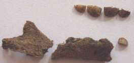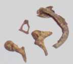


Left, Inhumation 6618; 2nd left, occipital bones; 2nd right, developing incisors; right, ear ossicles and tympanic ring
Well-preserved neonate, aged 28-34 weeks (premature).
The remains of a neonate (C6618) were recovered from a shallow pit in the foundations of the roundhouse, Timber Building 3. The child was lying on its left side in the foetal position. The following report summarises the results of a detailed study of the remains, the preservation and age of the individual.


Left, Inhumation 6618; 2nd left, occipital bones; 2nd right, developing incisors; right, ear ossicles and tympanic ring
The level of preservation of the human remains will affect the amount of information that can be collected. The hard clay and flint matrix made excavation difficult, and as a result only the upper half of the skeleton (skull, right arm and upper spine) was recovered. However, these bones were in an excellent state of preservation and in particular, the developing tooth crown, atlas, pars basilaris and even the ear ossicles were available for analysis.
It is not yet possible to assign a sex to non-adult remains, as the features used in this estimation appear at puberty. However, it is possible to attain a relatively precise age for foetal remains based on cranial and dental development. The age of the Silchester neonate was assessed using measurements of the pars basilaris (base of the skull), humerus and dental crowns (Fazekas and Kósa 1978; Scheuer and Black 2000; Schour and Massler 1941)
| Measurement (mm) | Estimated age | |
|---|---|---|
| Length of pars basilaris | 12.9 | 28 weeks |
| Distal humerus width | 13.2 | 34 weeks |
| Mineralisation of central and lateral maxillary incisors | n/a | 32 weeks-birth |
| Age range | 28-34 weeks | |
Full-term births are traditionally estimated to be between 38-40 weeks. At 34 weeks this neonate would have been 6 weeks premature and was probably not viable at birth. It was not possible to tell if the tympanic ring had already fused to the pars squama of the temporal bone, which normally occurs at birth.
Long bone fragments of two neonates, both full-term infants who died between 40-44 weeks.
One complete right femur and proximal fragments of the left femur, lefts and right tibiae and left and right humeri of a neonate. The maximum length of the right femur (86.62mm) indicates that the child was aged between 40-44 weeks (mean 42 weeks) at the time of death. Using the estimate already given above, this child was probably full-term (Scheuer and Black 2000). Such neonatal deaths are generally the result of internal (endogenous) factors caused by maternal illness, low birth weight or birth trauma, rather than being caused by the external environment into which they were born (Scott and Duncan 1999).


Left: Neonatal bones from 4774
Right: Neonatal femur from 4125
Distal half of a right neonatal femur. The distal aspect of the femur is too eroded for accurate measurements to be taken and the age cannot be ascertained. The bones are only slightly larger than those of 4774, and the child would have been of a similar age.
© Internet Archaeology
URL: http://intarch.ac.uk/journal/issue21/4/finds_human_bone.htm
Last updated: Wed Sept 12 2007